 The venous insufficiency pictures below are intended to aid in understanding the clinical evaluation of venous disease. There are several scoring systems physicians use to grade venous insufficiency. One of the most commonly used system is the CEAP classification. The C in the CEAP classification stands for clinical. What do the patients veins look like? E stands for etiology. What is the cause of the vein disease? A stands for anatomy. Which veins are involved? And finally, P stands for pathophysiology. Is the vein problem secondary to blockages or abnormal valves and reflux. Here is a picture guide to clinical section of the CEAP classification for venous insufficiency. All the following pictures are from patients seen at Lakeshore Veins, a varicose vein clinic in Mequon, Wisconsin
Images of Venous Insufficiency Stages
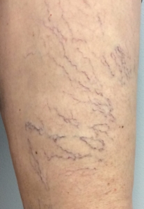
C1 disease – Spider veins or Telangiectasias
Spider veins are usually classified as the first level in the grading system, C1 disease. These veins are the small purple, blue or red vessels close to the skin. Additionally, spider veins are very common such that over half the US population has spider veins.
Also called telangiectasias, spider veins, occur in both women and men. They are much more common on the lower legs than anywhere else on the body.
Many patients seek care for spider veins or telangiectasias because they do not like the appearance. This young woman sought care at Lakeshore Veins for the spider veins on her left thigh. These veins had no associated symptoms and were treated with sclerotherapy to improve the cosmetic appearance.
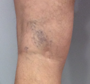
However, not all telangiectasias are treated for cosmetic reasons. This patient had underlying venous insufficiency and developed spontaneous bleeding. She noted a large amount of blood on the bathroom floor after stepping out of the shower. No trauma occurred in the area. Spontaneous bleeding after a shower can occur because the warmth of the shower causes the vessels to dilate and the skin to soften. Spider vein treatment can be covered by insurance if symptoms are present. It is important to seek medical advice if you have symptoms.
C2 disease – Venous insufficiency with Varicose Veins
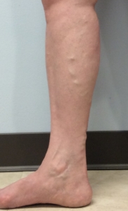
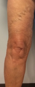
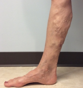
Varicose veins are the large bulging veins under the skin usually in the legs. Often, varicose veins are symptomatic causing heavy, tired, sore, itchy legs. But, they can also be have no associated symptoms. Once present, varicose veins will not usually go away. However, varicose veins are not usually dangerous and usually only require treatment if symptoms are present.
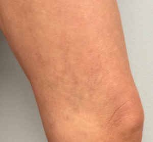
Varicose veins can occur in the thigh as in this picture. Although, they can be present in the lower leg or both.
Varicose veins can occur in young patients as early as their teens. Additionally, varicose veins can be present in active patients with a normal body weight. Generally, there is a strong genetic cause to varicose veins. Therefore, you are much more likely to have varicose veins if your mother or father has varicose veins.
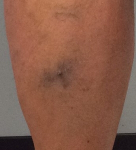
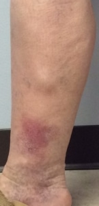
Sometimes the varicose veins can clot and cause pain. Note the redness in the calf of this patient who also noted a hard lump. Clotted superficial veins are called superficial thrombophlebitis or phlebitis for short. Mild phlebitis will usually resolve after several weeks. However, it is important to seek medical attention if the area of phlebitis is larger than 5 cm in size, is located in the upper thigh, or is getting larger. Additionally, consult a physician if you have any medical conditions that makes your blood clot abnormally (cancer, recent pregnancy, immobility, recent surgery). Patients with recurrent phlebitis should have an ultrasound to evaluate for an anatomical cause of the phlebitis.
C3 disease – Venous insufficiency with swelling
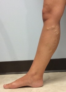
As venous insufficiency progresses, swelling is seen. Swelling usually starts at the ankle but can include the entire leg. In this patient, note the varicose veins in the calf. Also, there is leg swelling with sock lines seen at the ankle and the knee. Classically, leg swelling caused by venous insufficiency is worse at the end of the day and better in the morning. Additionally, leg swelling can be worse after travel. Both conservative care including use of graduated compression stocking as well as treating the underlying venous insufficiency can help with leg swelling.
C4 – Venous insufficiency with skin changes
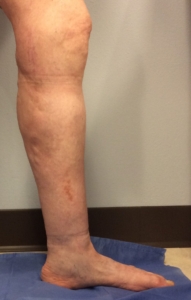
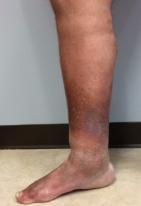
As venous insufficiency progresses further, the skin becomes affected. Note the slight brown discoloration around the ankle and lower leg. This brown discoloration is a very early sign of skin changes. The distended veins leak slightly and the hemosiderin in the blood stains the skin. The skin staining is usually permanent.
This image shows a leg with much worse skin changes and with leg swelling. As the skin changes progress the lower leg becomes browner, the skin drier and more fragile. It is important to treat the underlying venous insufficiency before wounds develop.
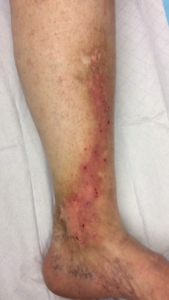
In addition to the brown skin changes, patients can also develop red, inflamed, scaly itchy skin known as venous eczema. Topical treatments are helpful; however, these patients do require treatment of their venous insufficiency to control their symptoms.
Another picture of a Lakeshore Veins patient with venous insufficiency skin changes and a large bulging vein above the area of red, scaly, painful skin.
C5 – Venous insufficiency with healed ulcer
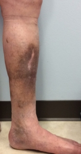
There are extensive venous stasis skin changes on the left leg with brown discoloration. The whiter region in the center of the brown are is in the region of the patients healed venous stasis ulcer. He was treated with ablation of his insufficient Great Saphenous vein and was able to heal a long-standing ulcer. Also, his ulcer has remained healed for several years.
C6 – Venous insufficiency with active ulceration
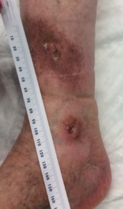
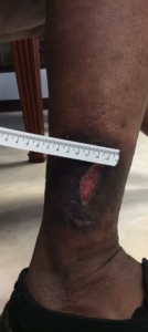
C6 is the worst classification of chronic venous insufficiency. In this category, there is an active open venous ulcer on the skin.
Active venous insufficiency ulcers are often on the lower leg close to the ankle but can occur anywhere usually below the knee. Venous insufficiency ulcers are difficult to heal. Additionally, the ulcers often are very moist and will soak through dressings. Therefore, treating the under lying venous insufficiency is very helpful in preventing new ulcer from developing.
Venous Insufficiency Picture Summary
The picture guide of the clinical classification of venous insufficiency shows how the vein disease is categorized from the mildest to the most severe disease. There are many available treatments to address venous insufficiency. Therefore, it is important to discuss any symptoms or concerns with a health care provider or vein specialist near you.
